
In the news bulletin of ANT1, Nikos Maratheftis, neurosurgeon
The neurosurgeon Nikos Maratheftis appeared in the ANT1 news bulletin, and spoke about the spine surgeries only with local anesthesia that were performed for the first time in Greece at the Euroclinic in Athens .
Disc hernia: new data in its treatment
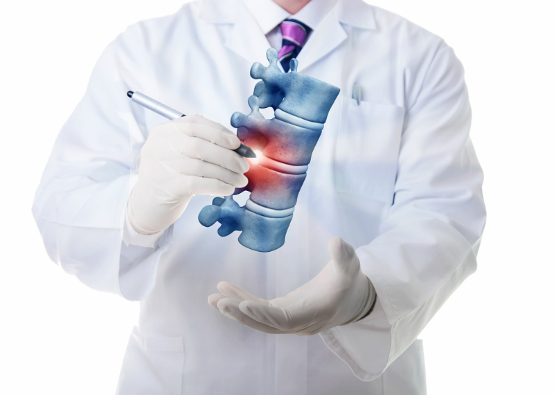
In recent years, spinal surgery has made tremendous progressUntil recently, or even at the present time, when a patient learned that they needed back surgery, they experienced great fear and uncertainty. Everyone has a friend, acquaintance or neighbor who, after a back surgery for a herniated disc, suffered for a long time, did not solve […]
Percutaneous spine surgeries
In modern surgery, with the help of technology and the accumulation of experience, new methods have been developed, which help surgeons perform operations that in the past were particularly traumatic, with minimal risk of surgical trauma. A typical example is spine surgeries, such as spinal fusion. Percutaneous spine surgeries significantly reduce patients’ […]
Pain Treatment with Radiofrequency Electrode Application
Radiofrequency therapy is indicated for the treatment of neuropathic pain, neuralgia, but also persistent musculoskeletal pain such as sciatica, low back pain and neck pain, which is continuous, has an anatomically clear distribution and is resistant to conservative treatment (medicines, patches, etc.). Neuropathic pain can be treated with the use of radiofrequency. Examples […]
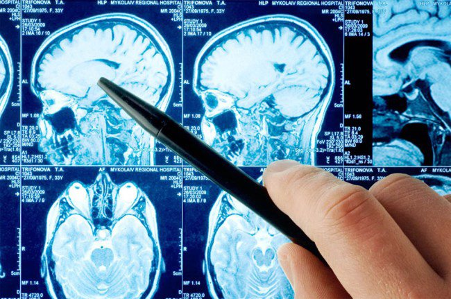
Postoperative chronic pain
This category includes a number of conditions such as the so-called failed back / failed neck pain syndrome. Many of these patients continue to suffer from pain, although clinical and imaging tests (X-rays, MRI scans, etc.) do not identify any cause of their pain. Implantation of spinal cord stimulators in the spine In […]
Tumours
Tumours of the spine, spinal cord and nerves